
Introduction:
The lungs are essential organs in the chest, responsible for breathing and gas exchange. They take in oxygen from the air and release carbon dioxide, a waste product of metabolism. This exchange is crucial for maintaining cellular function and overall health. All body cells require oxygen to produce energy. They must also eliminate carbon dioxide to prevent toxicity.
Location of the Lungs:
The lungs are situated within the thoracic cavity, a protected area formed by the rib cage and other structures. This space is vital as it provides a secure environment for the lungs. It allows them to expand and contract during breathing. Additionally, it supports them structurally.
Boundaries of the Thoracic Cavity:
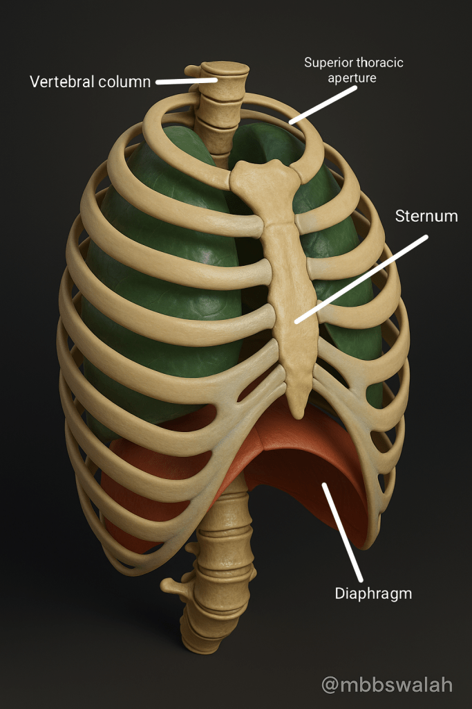
Top:
The superior thoracic aperture is also known as the thoracic inlet. It is the opening at the top of the thoracic cavity. This opening allows critical structures, such as the trachea (windpipe) and esophagus, to enter and exit.
Bottom:
The diaphragm is a muscular sheet that separates the thoracic cavity from the abdominal cavity. During inhalation, the diaphragm contracts and flattens, creating a vacuum that pulls air into the lungs.
Front:
The sternum, or breastbone, lies vertically in the center of the chest. It provides a protective wall for the heart and lungs.
Back:
The thoracic spine consists of vertebrae that encase the spinal cord and provide support and flexibility to the upper body.
General Characteristics:
Lung tissue is spongy and elastic, allowing it to expand and contract with each breath. Their conical shape makes sure the arrangement maximizes the surface area for gas exchange. Oxygen is absorbed, and carbon dioxide is expelled.
Gross Anatomy: External Features and Internal Divisions
1. Lobes and Fissures:
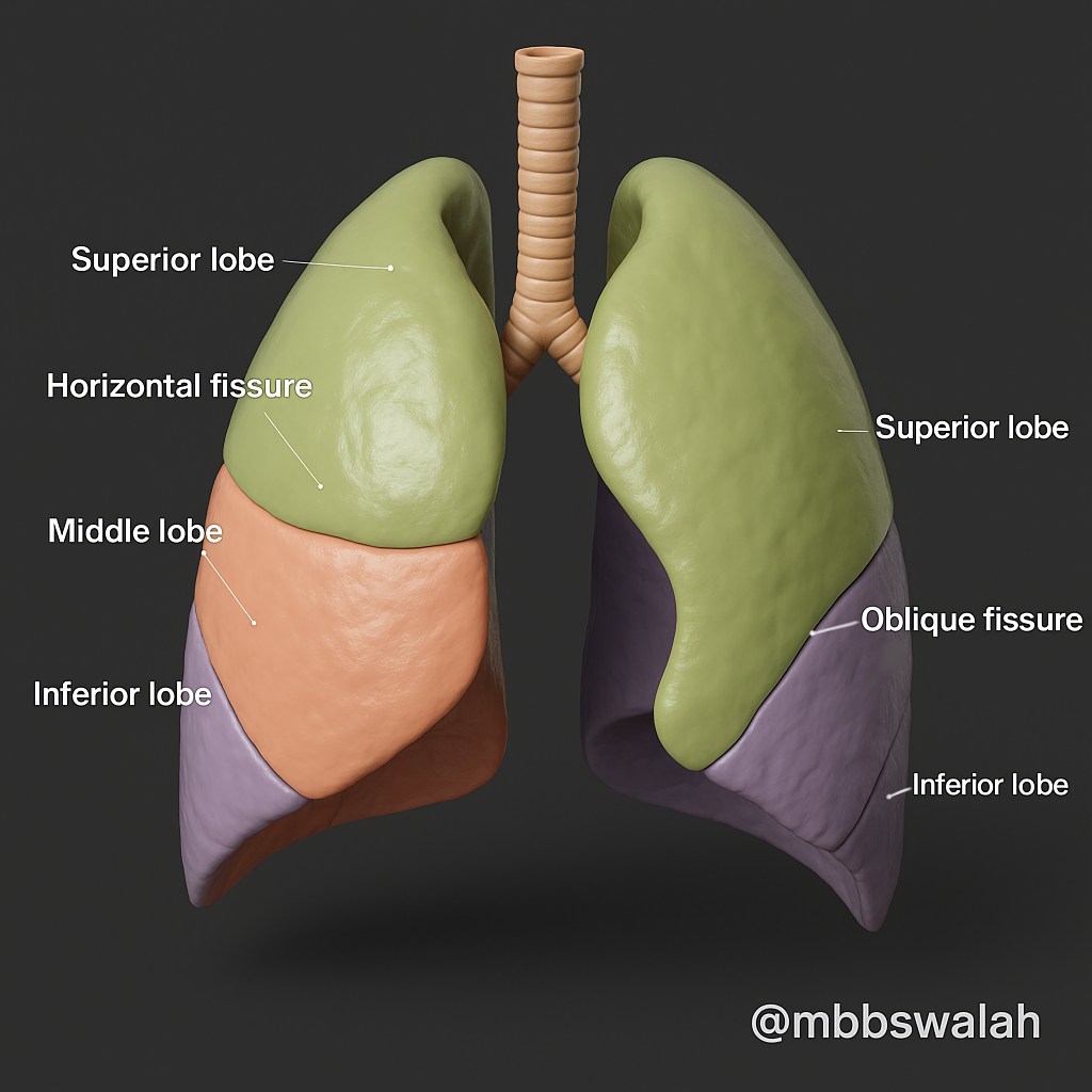
The lungs are divided into lobes by fissures:
– The right lung has three lobes (upper, middle, lower) divided by horizontal and oblique fissures.
– The left lung has two lobes (upper and lower) divided by an oblique fissure. This segmentation is important for understanding lung mechanics. It is also useful for medical procedures. These procedures allow for targeted surgical interventions while preserving maximum lung function.
2. Surfaces:
Costal Surface:
The largest surface, it has a smooth, convex shape fitting against the ribcage, aiding in expanding when breathing.
Mediastinal Surface:
This concave surface faces the center of the chest (mediastinum). It includes the hilum. Important vessels and air passages enter and exit the lungs through the hilum.
Diaphragmatic Surface:
The base of the lung rests on the diaphragm and is concave, allowing space for the diaphragm’s movement during respiration.
3. Apex and Base:
Apex:
The uppermost part of the lung extends slightly above the first rib. This is critical for ensuring that the lung’s top portion is adequately positioned for air intake.
Base:
The bottom part of the lung rests on the diaphragm. It is where the lung interfaces with the muscular structure. This structure aids in breathing.
4. The Hilum:
The hilum of each lung serves as the entry and exit point for several critical structures:
Left Hilum: Contains a single main bronchus, which branches off to supply the left lung. The arrangement here is important. It allows for the unique layout of the left lung. This layout makes space for the heart and the aorta.
Right Hilum: Contains three main bronchi (one for each lobe), along with the pulmonary arteries and veins. The right main bronchus is wider and more vertical. This makes it easier for inhaled objects to enter this side of the lung. This scenario is a common clinical consideration.
Understanding the structure and function of the lungs is crucial for diagnosing and treating respiratory diseases effectively. This includes their lobular organization and the significance of the hilum.
This knowledge helps in surgeries, managing lung infections, and understanding lung cancer’s impact on respiratory health.


Leave a comment