
The Four Chambers (Rooms Inside)
- The heart’s inside is divided into four distinct rooms, each with a specific role.
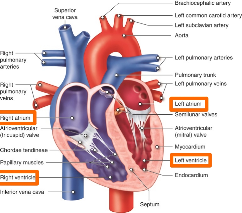
- Right Atrium: Receives used blood from the body. It gets this blood through three veins. The first is the superior vena cava, which carries blood from the upper body.
- The second is the inferior vena cava, which carries blood from the lower body.
- The third is the coronary sinus, which serves the heart muscle itself. Inside, there’s a shallow spot called the fossa ovalis, which is a remnant of a fetal heart opening. There are also openings for the vena cavae and coronary sinus. Muscular ridges, called pectinate muscles, help it contract.
- Right Ventricle: The right ventricle gets used blood from the right atrium through the tricuspid valve. It pumps the blood to the lungs to get oxygen. Blood leaves through the pulmonary valve into the pulmonary artery.
- Inside, the walls have ridges (trabeculae carneae). Cone-shaped muscles (papillary muscles) are connected to the tricuspid valve by strong strings called chordae tendineae. These prevent the valve from flipping backward when the ventricle squeezes. A muscle band called the moderator band helps coordinate contraction.
- Left Atrium: Receives fresh, oxygenated blood from the lungs via the pulmonary veins (usually four). Its inner walls are mostly smooth, except in a small pouch called the left auricle. It pumps this fresh blood into the left ventricle through the mitral (or bicuspid) valve.
- Left Ventricle: The left ventricle gets fresh blood from the left atrium. It has the thickest walls because it pumps blood with strong force to the whole body. Blood flows out through the aortic valve into the aorta. Like the right ventricle, it has muscle ridges (trabeculae carneae). It also has cone-shaped muscles (papillary muscles). These muscles are connected to the mitral valve by strong strings (chordae tendineae). The part leading to the aorta is called the aortic vestibule.
- The differences in chamber structure match their jobs: atria are receiving rooms (thinner walls), ventricles are powerful pumps (thicker walls).
Heart Valves (One-Way Doors)
- Four valves ensure blood flows in only one direction, preventing backflow.
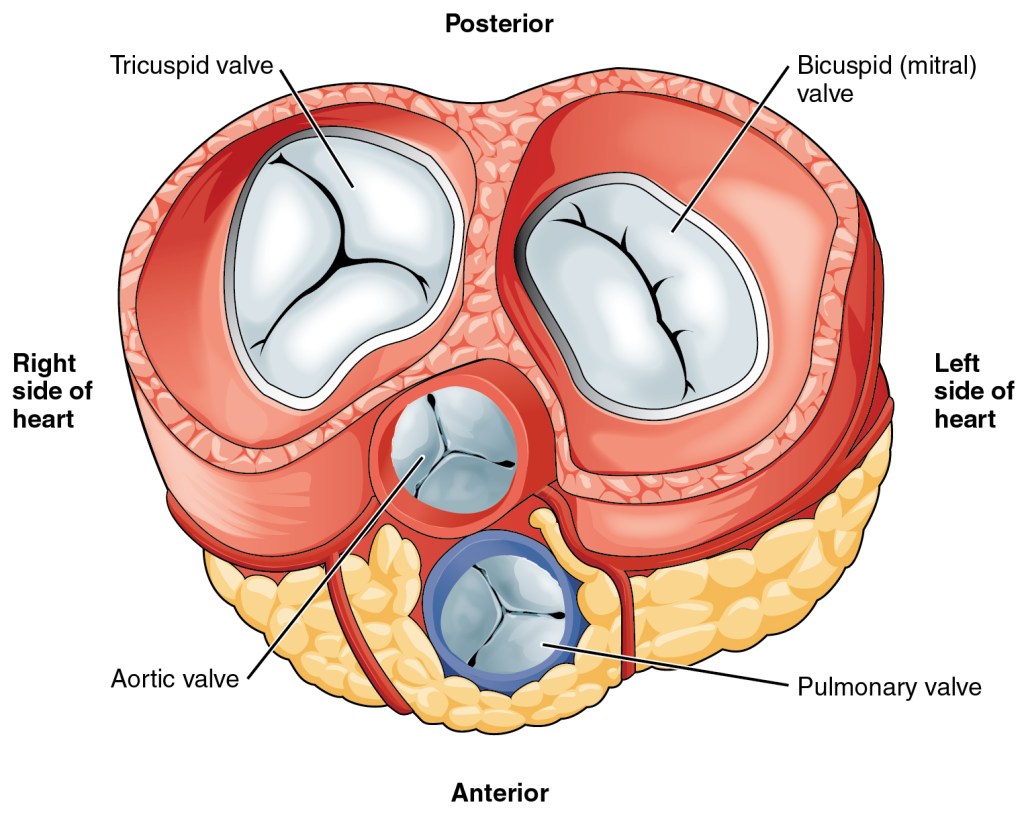
- Tricuspid Valve: Between the right atrium and right ventricle. Has three flaps (leaflets/cusps). Opens to let blood into the ventricle, closes to prevent backflow into the atrium when the ventricle pumps.
- Pulmonary Valve: Between the right ventricle and pulmonary artery. Has three half-moon shaped cusps. Opens when the ventricle pumps blood to the lungs, closes to prevent backflow into the ventricle when it relaxes.

- Mitral Valve (Bicuspid Valve): The mitral valve is between the left atrium and left ventricle. It lets blood flow down to the ventricle and stops it from going back up. Has two flaps (leaflets). Opens to let blood into the ventricle, closes to prevent backflow into the atrium when the ventricle pumps.
- Aortic Valve: Between the left ventricle and the aorta. Also has three half-moon shaped cusps. Opens when the ventricle pumps blood to the body, closes to prevent backflow into the ventricle when it relaxes.
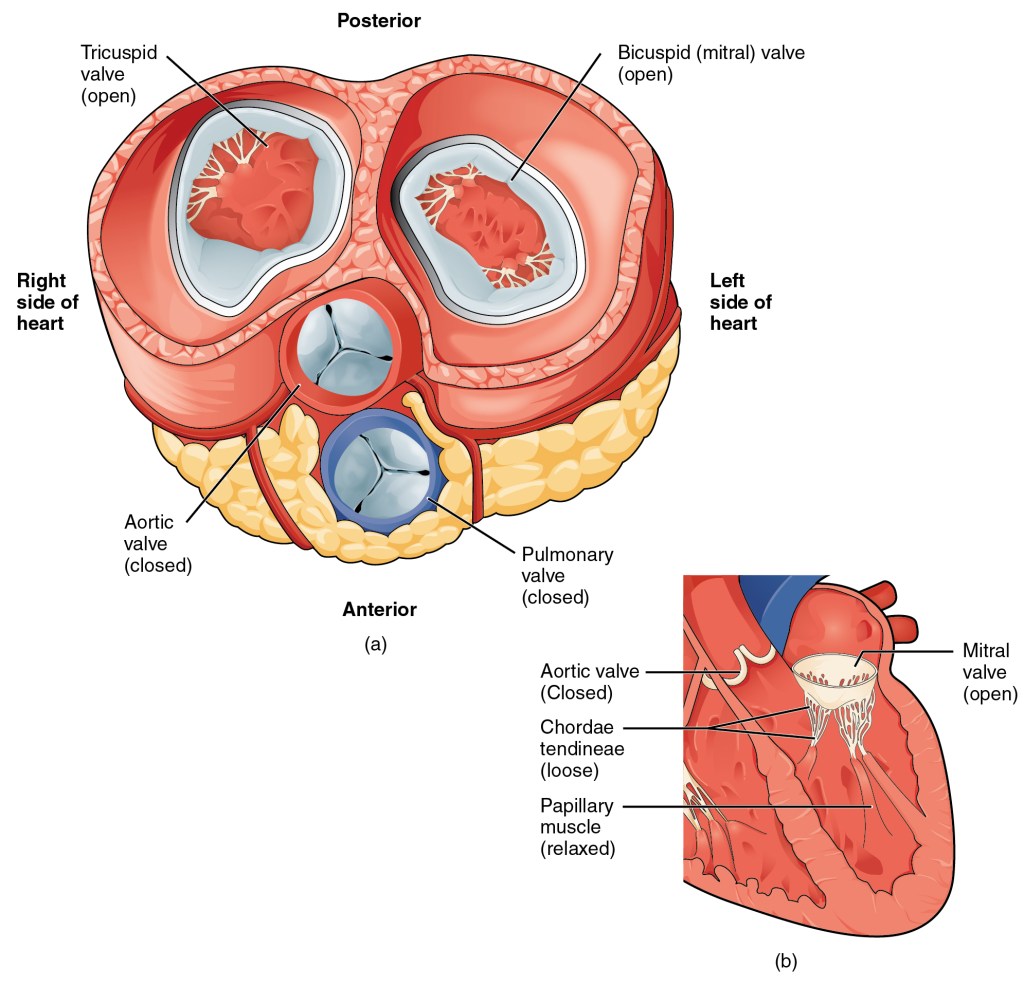
- The coordinated opening and closing of these valves with the heart’s pumping is key for efficient one-way blood flow.
Septum (The Dividing Walls)
- Muscular walls called septa divide the heart into left and right sides.
- Interatrial Septum: The interatrial septum is a thin wall. It separates the right and left upper chambers of the heart (atria). Contains the fossa ovalis (remnant of fetal circulation bypass). Its main job in adults is to keep used blood (right) from mixing with fresh blood (left).
- Interventricular Septum: The interventricular septum is a thick wall. It separates the right and left lower chambers of the heart (ventricles). Its thickness is needed to handle the high pressure from pumping ventricles. It has a big muscle part and a small thin part made of membrane at the top. It prevents mixing of used and fresh blood in the ventricles and contains important parts of the heart’s electrical system.
- These walls are essential for keeping the lung circulation (pulmonary) separate from the body circulation (systemic), ensuring efficient oxygen delivery.
The Flow of Blood Through the Heart (The Journey)
- Blood flow follows a precise path ensuring oxygenated blood reaches all tissues.
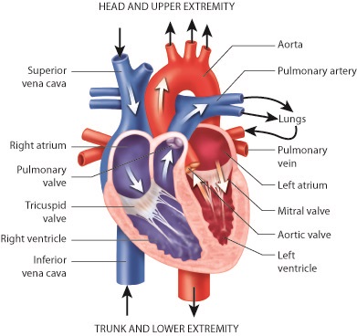
- Used blood enters Right Atrium: Blood low in oxygen comes back from the body. It returns via the superior and inferior vena cava into the right atrium.
- Flows to Right Ventricle: Blood passes through the tricuspid valve into the right ventricle.
- Right Ventricle Pumps to Lungs: The right ventricle pumps the used blood. It moves the blood through the pulmonary valve into the pulmonary artery. The blood is heading to the lungs.
- Fresh blood Returns to Left Atrium: In the lungs, blood drops off carbon dioxide and picks up oxygen. This fresh blood comes back through the pulmonary veins into the left atrium.
- Flows to Left Ventricle: Blood passes through the mitral valve into the left ventricle.
- Left Ventricle Pumps to Body: The powerful left ventricle pumps the fresh blood through the aortic valve. The blood then flows into the aorta, which sends it to the rest of the body.
- This cycle keeps going. The right side handles used blood, and the left side handles fresh blood. This ensures efficient oxygen delivery. The chambers and valves work together perfectly to keep blood moving one way. Atria receive blood; ventricles pump blood. Valves act as crucial one-way gates.
Layers of the Heart Wall (The Heart’s Construction)
- The heart wall has three distinct layers.

- Pericardium (Outermost): A double-layered sac.
- Outer fibrous layer: Tough, inelastic connective tissue; protects the heart, anchors it, prevents overfilling.
- Inner serous layer: Has two parts. Parietal lines the fibrous layer. Visceral, also called epicardium, sticks directly to the heart muscle.
- Pericardial cavity: Space between serous layers with lubricating fluid to reduce friction during heartbeats.
- Myocardium (Middle): The thickest layer, made of heart muscle tissue (cardiomyocytes). This is the muscle that pumps blood. Its thickness varies; it’s thickest in the left ventricle because it works hardest. Contains the electrical system and gets blood from coronary arteries.
- Endocardium (Innermost): Thin layer lining the chambers and covering the valves. Made of smooth endothelium (like blood vessel lining). Its smoothness prevents blood clots from forming inside the heart. Below the endothelium is connective tissue with tiny blood vessels, nerves, and electrical fibers (Purkinje fibers).
The Heart’s Electrical Conduction System (The Spark Plugs)
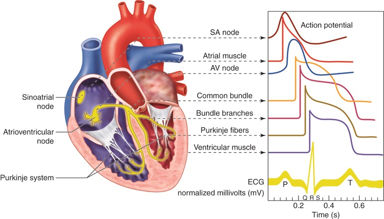
- The heart beats because of a special electrical system that generates and sends signals through the heart muscle, coordinating its contractions.
- Sinoatrial (SA) Node: Located in the upper right atrium wall. It’s the heart’s natural pacemaker, starting electrical signals regularly (usually 60-100 times per min.
- Signal Spreads: The signal spreads across the atria, making them contract.
- Atrioventricular (AV) Node: Located lower in the right atrium near the tricuspid valve. It delays the signal slightly, ensuring the atria empty before the ventricles pump.
- Bundle of His: The signal travels from the AV node down this bundle of fibers. It is located in the wall between the ventricles (interventricular septum).
- Bundle Branches: The Bundle of His splits into right and left branches going towards each ventricle down the septum.
- Purkinje Fibers: These branches spread out into a network of fine fibers throughout the ventricle walls (myocardium). They deliver the signal rapidly for efficient ventricle contraction.
- Control: The body’s autonomic nervous system can adjust the SA node’s rate, changing the heart rate based on body needs.
Conclusion
- The human heart’s anatomy is a marvel, with complex structures working together for efficient blood pumping.
- Its location, chambers, vessels, valves, septum, and wall layers all contribute to its vital life-sustaining role.
- The heart’s electrical system ensures a rhythmic beat for proper blood pressure and tissue supply.
- While sharing basics with other mammals, the human heart has unique features shaped by evolution and specific human needs.


Leave a comment