
Introduction
- The human heart is the main engine for our blood circulation system, located near the center of our chest.
- Its complex structure and coordinated work are crucial. They send oxygen and nutrients to all our body cells. They also take away waste.
- Knowing how the heart is built helps us understand how it keeps us healthy. It also helps us understand what goes wrong in heart diseases.
- This guide will give you a detailed look at the heart’s outside (external) and inside (internal) parts. It will include its location, rooms (chambers), major blood pipes (vessels), doors (valves), and walls (septum). You’ll also learn about its layers, electrical system, and how it compares to hearts in other animals.
- This information is aimed at those interested in the details of how the human body works.
External Anatomy (The Outside of the Heart)
Where is the heart?
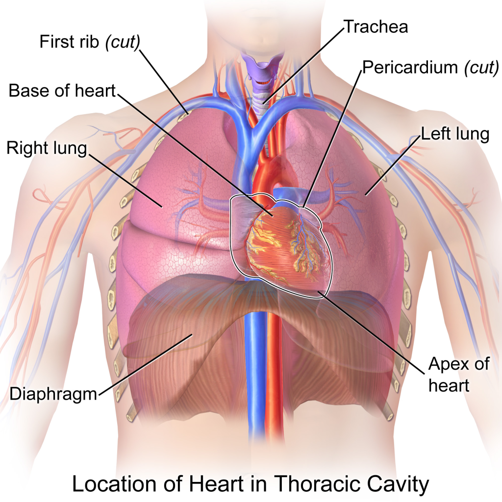
- The heart sits in your chest cavity, in an area called the middle mediastinum, right between your lungs.
- It’s not perfectly centered; about two-thirds of it is on the left side of your body’s middle line.
- It’s located behind your breastbone (sternum) and nearby rib cartilages. It is in front of crucial structures. These include your food pipe (esophagus), the main artery going down your chest (descending thoracic aorta), and your spine.
- How is the Heart Positioned?
- The top part is called the base. It is where the big blood vessels connect. These vessels are located around the level of our third rib cartilage.
- The bottom tip, the apex, points down. It also points forward (antero-inferiorly) and to the left. This is usually around the space between your fifth and sixth ribs.
- The slant causes the heart to sit diagonally in the chest. Its right side is more towards the front. The left side is more towards the back.
- The apex pointing left makes a little dent in the left lung called the cardiac notch.
- The whole heart is wrapped in a protective bag called the pericardium.
- This specific location and angle are important for how the heart works with the lungs and major blood vessels. It also helps doctors listen to our heart or take images.
- Chambers (Rooms You Can See from Outside)
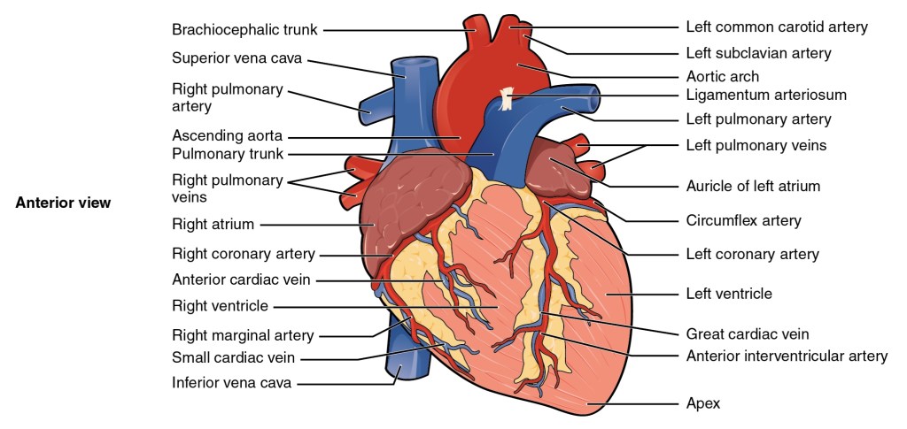
- The heart has four rooms. Two upper ones are called atria: right and left. Two lower ones are called ventricles: right and left.
- From the outside, a groove called the coronary sulcus (or atrioventricular groove) shows where the atria and ventricles meet.
- The front surface of the heart is mostly made up of the right atrium and right ventricle.
- The left ventricle forms the heart’s tip (apex). It forms most of the left edge. It also forms a big part of the bottom surface.
- The left atrium is mostly at the back and forms the base.
- Jobs of the Chambers (External View Related)
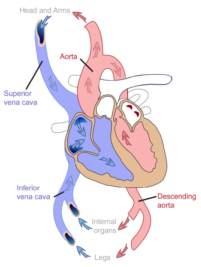
- The right atrium receives used blood that is low in oxygen. This blood comes back from the body via the superior and inferior vena cava.
- The left atrium takes in fresh blood (rich in oxygen) coming from the lungs through the pulmonary veins.
- The right ventricle pumps the used blood to the lungs via the pulmonary artery.
- The left ventricle pumps blood to the rest of the body through the aorta.
- Major Blood Vessels (Pipes Connected to the Heart)
- Several large blood pipes connect directly to the heart to move blood in and out.
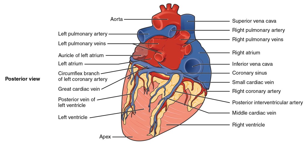
- Aorta: The aorta is the largest artery. It starts from the left side of the heart and carries oxygen-rich blood to the entire body. We can see its beginning parts (ascending aorta, arch, start of descending aorta) at the heart’s base. The heart’s own fuel lines, the coronary arteries, branch off from the very start of the aorta.
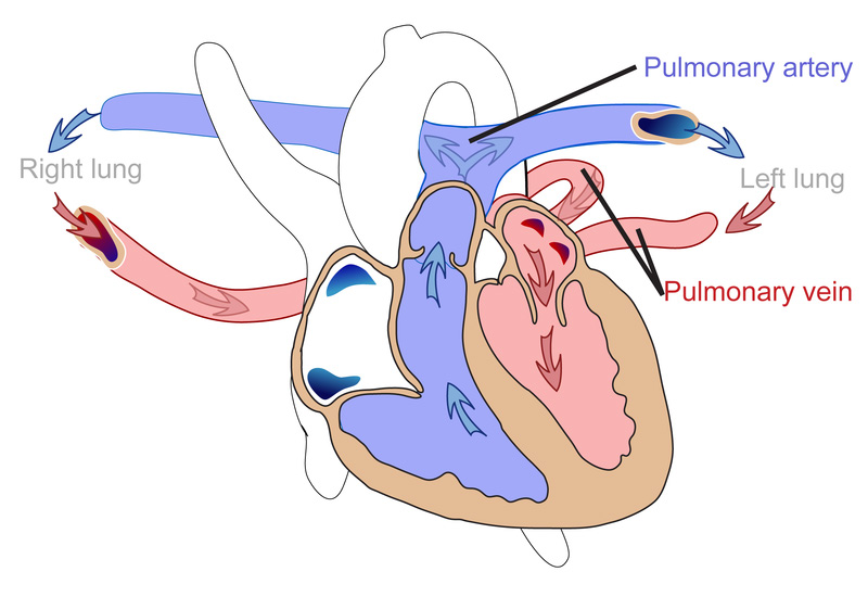
- Pulmonary Artery (Trunk): The pulmonary artery starts from the right side of the heart and carries used blood to the lungs to pick up oxygen. We can see where it splits into left and right pulmonary arteries on the heart’s surface.
- Vena Cava (Superior and Inferior): These are large veins. The superior vena cava (SVC) brings used blood from the upper body to the right atrium, while the inferior vena cava (IVC) brings used blood from the lower body to the right atrium.
- Pulmonary Veins: Pulmonary veins carry oxygenated blood from the lungs to the left side of the heart. We can see where they enter the back of the left atrium.
- These big vessels are the main highways for blood traveling between the heart and the body, allowing oxygen and carbon dioxide exchange.
- Surfaces and Borders (Shape of the Heart)
- The outside of the heart has different faces or surfaces:
- Anterior (front/sternocostal): Mostly the right ventricle.
- Posterior (back): Forms the base, mostly the left atrium.
- Inferior (bottom/diaphragmatic): Made of both left and right ventricles.
- Right pulmonary: Made of the right atrium.
- Left pulmonary: Mostly the left ventricle.
- The edges or borders define these surfaces:
- Right border: Right atrium.
- Inferior border: Part of the right ventricle left ventricle.
- Left border: Mainly the left ventricle and a bit of the left atrium.
- Superior border: Both atria and the large blood vessels.
- Knowing these helps describe the heart’s location and size accurately, especially in medicine.
- External Sulci (Grooves on the Outside)
- The heart’s surface has grooves (sulci) that mark where the chambers are divided inside and hold the heart’s own blood vessels (coronary vessels).
- Coronary Sulcus (Atrioventricular Groove): A deep groove going around the heart, separating the atria above from the ventricles below. It holds important coronary arteries and veins.
- Anterior Interventricular Sulcus: On the front surface, running down towards the apex. It marks the division between the right and left ventricles on the front. It contains the main front coronary artery (LAD) and a major vein.
- Posterior Interventricular Sulcus: On the back surface, also running down towards the apex. It marks the division between the ventricles on the back and contains the posterior coronary artery and another major vein.
- These grooves are like landmarks showing the chambers underneath and the paths of the vessels feeding the heart muscle.
- Functions of External Parts
- The outside structure of the heart is built for efficiently pumping blood throughout the circulatory system.
- Its location in the chest protects it and allows it to work closely with the lungs and major blood vessels.
- The major vessels are the entry and exit routes for blood, ensuring oxygen delivery and waste removal.
- The surfaces and borders define its shape and relationship with nearby organs within the chest.
- The grooves show chamber divisions and hold the vital coronary vessels that nourish the heart muscle.
- The heart’s size is about as big as your fist. This size is suited for its space in the chest. It is also suited for its crucial job.


Leave a comment