
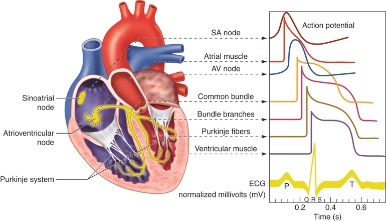
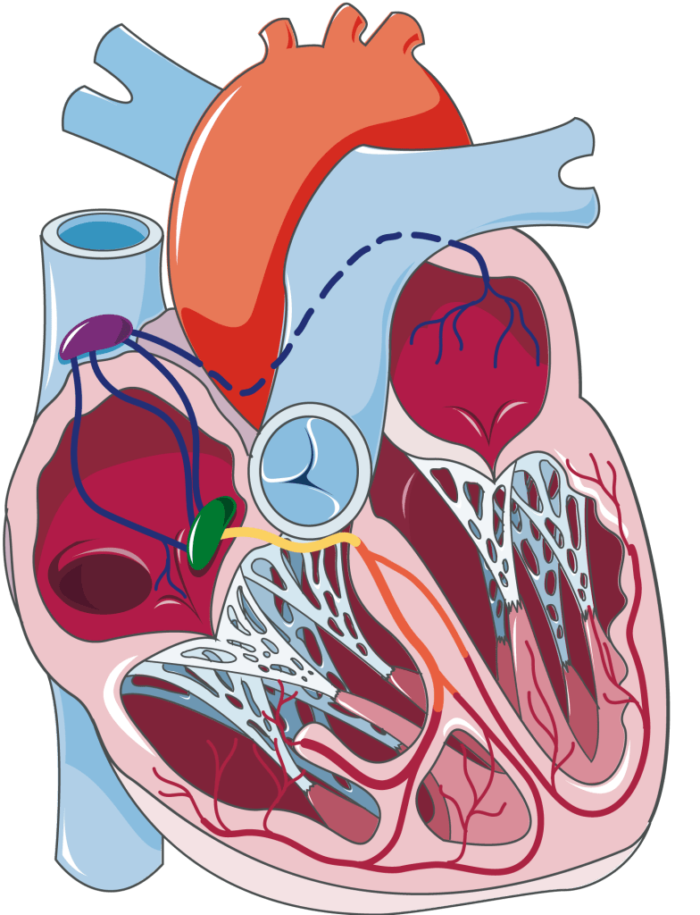
The conducting system of the heart consists of specialized cardiac muscle cells. They generate and transmit electrical impulses. This process ensures the heart beats in a coordinated and rhythmic manner. The main components are:
Sinoatrial Node
The sinoatrial node is the primary pacemaker in mammalian heart.
It is situated in the right atrium close to the opening of the superior vena cava.
It is about 1.5 cm long and 0.5 cm wide in human beings.
It contains the pacemaker (P) cells and few myofilaments.
The P cells generate the impulse, which is transmitted in the conducting system for excitation and contraction of heart muscles.
The action potentials generated in SA node are of slow response type.
The velocity of conduction of impulse in SA node is slow (0.05 m/s).
Internodal Pathways
There are three internodal pathways that connect SA node and AV node.
1. The anterior internodal pathway is called the tract of Bachman. The middle internodal pathway is called the tract of Wenckebach. The posterior internodal pathway is called the tract of Thorel.
2. The internodal pathways merge into the AV node.
3. The velocity of conduction of impulse in internodal pathways is about (1 m/s).
4. From SA node, a conducting tract arises and directly enters into the left atrium. This is called interatrial tract or Bachman’s bundle.
Atrioventricular Node
The atrioventricular node is located in the lower part of the right atrium. It is close to the interatrial septum and just above the atrioventricular ring.
It is 22 mm in length, 10 mm in width and 3 mm in thickness.
In AV node, the fiber diameter is small and there are multiple sub-branches. Therefore, the rate of impulse conduction is slow in AV node (0.05 m/s).
Usually, a delay of about 0.1 second occurs for the impulse to be transmitted through AV node. This is called AV nodal delay.
This delay is shortened by sympathetic stimulation and lengthened by parasympathetic stimulation to the heart. The weak impulses may even die out in AV node.
The AV node can slow down the transmission of rapid impulses from the SA node to the ventricle. This ability is also called decremental conduction.
The action potentials generated in AV node are of slow response type.
Pacemaker cells are also present in the AV node. However, the AV node is not normally the pacemaker. The rate of impulse formation in the AV node is lower than that of the SA node. The (P) cells of AV node are suppressed by the SA nodal impulses.
However, when SA node stops producing impulses, AV node becomes the pacemaker of the heart.
HIS Bundle
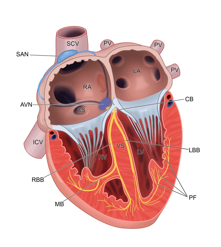
This is a small bundle of fibers that arises from AV node and terminates in the Purkinje system.
This is situated below the AV node and passes towards the interventricular septum.
The fibers are present in the form of a bundle. This is called bundle of His. It is not called the bundle of Her, as it was described by W His in 1893.
The length of His bundle is about 1 cm. Upon entering the interventricular septum, it divides into the right bundle branch and the left bundle branch.
When SA node and AV node are defunct, the bundle of His generates impulses.
Bundle Branches
His bundle divides into the right bundle branch (RBB) that conducts impulses to the right ventricle. It also divides into the left bundle branch (LBB) that conducts impulses to the left ventricle. The bundle branches enter the ventricular walls and then branch out into very small bundles of fibers in the inner walls of the
ventricular muscle. These fibers are termed as Purkinje fibers. Bundle branches also have the potentiality to generate impulses.
Right Bundle Branch
Right bundle branch is longer and thinner than the left bundle branch. It exclusively supplies right ventricle.
Left Bundle Branch
Left bundle branch bifurcates into two divisions. The anterior division supplies the anterior portion of the left ventricle. The posterior division supplies the posterolateral portion of the left ventricle.
Purkinje Fibers
This is a network of small bundles of conducting fibers. They are present throughout the subendocardial regions of both the right and left ventricles.
The cells of the Purkinje system (are also called Purkinje cells) are the largest cells in the heart.
Numerous gap junctions (low impedance electrical synapses) are present between the cells.
The fiber has larger diameters. It features low impedance cell-to-cell connections. Because of these characteristics, the rate of impulse conduction is highest in the Purkinje fibers.
The conduction rate is almost 4 m/s.
The action potentials generated in the Purkinje fibers are of fast response type. They resemble those produced in the ventricular muscles.


Leave a comment