
The fetal circulatory system is a special design that helps a baby grow inside the womb. It works differently from a newborn’s system.
It sends blood in unique pathways. This ensures the fetus receives oxygen and nutrients from the mother until birth. This intricate network of vessels and specialized structures differs significantly from the postnatal circulatory system.
The main reason is that the fetus relies entirely on the placenta for gas exchange and nutrient acquisition. Fetal circulation is a specialized system. It transports oxygen and vital nutrients from the mother’s bloodstream to the fetus.
This supports its growth and development. At the same time, it collects carbon dioxide and other waste products produced by the fetus. These are directed back to the mother’s circulation, where they are processed and eliminated.
This unique system allows the fetus to thrive in the womb without relying on its own lungs. It also does not rely on its liver or kidneys. These are not yet fully functional. This is done by a sophisticated system. It involves the placenta and the umbilical cord. Specialized circulatory shunts allow blood to bypass the non-functional fetal lungs and liver.
The main heading to be covered to understand fetal circulation:
- Key Difference between Fetal and Adult Circulatory Systems.
- The Placenta and Umbilical Cord: Essential Components.
- Fetal Shunts: Bypassing Non-Functional Organs.
- The Path of Oxygenated and Deoxygenated Blood.
- Circulatory Changes at Birth: Transition to Neonatal Circulation.
Key difference between fetal and adult circulation:
At birth, the circulatory system undergoes major changes to help the newborn survive independently. As the baby takes its first breath, the lungs expand. Blood flow is redirected away from the fetal shunts toward the lungs and liver. Temporary structures like the foramen ovale and ductus arteriosus begin to close. This closure allows the baby’s heart and blood vessels to function normally. They now follow the adult pattern for oxygen and nutrient delivery.
Site of gas exchange :
In the fetus, this crucial function is performed by the placenta. Oxygen and nutrients from the mother’s blood are transferred to the fetal circulation. Waste products are moved in the opposite direction. This necessitates a completely different circulatory arrangement in the fetus to bypass the fluid-filled, non-functional lungs.
Specialized shunts :
To ensure efficient delivery of oxygenated blood to the developing body, the fetal circulatory system employs several specialized shunts . These include :
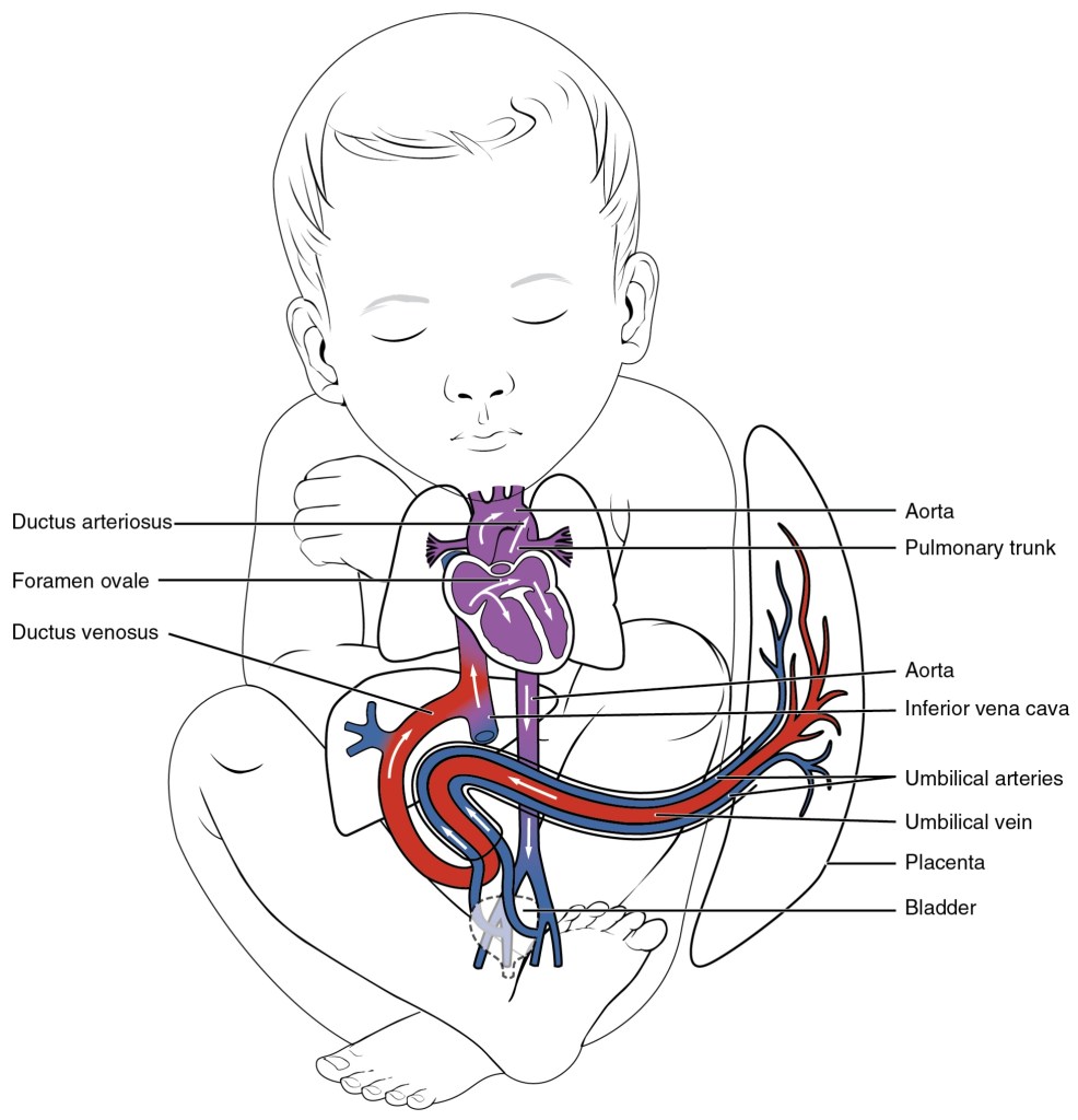
- Ductus venosus :This is a special fetal blood vessel. It channels oxygen-rich blood from the placenta directly into the inferior vena cava. This allows most of the blood to bypass the liver. This shortcut ensures that highly oxygenated blood quickly reaches the heart and vital organs. This supports the rapid growth and development of the fetus.
- Foramen ovale: an opening between the right and left atria of the heart. It permits oxygenated blood to bypass the right ventricle and lungs. The blood flows directly into the left atrium.
- Ductus arteriosus : a blood vessel connects the pulmonary artery to the aorta. It shunts blood away from the lungs and into the systemic circulation.
These shunts are crucial for fetal life but close shortly after birth, becoming ligamentous remnants in the adult circulatory system.Their closure marks a critical step in the transition to independent breathing and circulation.
Blood flow pathway :
Fetal circulation involves parallel circuits. Oxygenated blood from the placenta is directed to the systemic circulation, particularly the brain and heart. This process bypasses the lungs and
liver. In the fetus, this parallel arrangement ensures the most oxygenated blood reaches the vital organs. These organs have the highest metabolic demands. It supports their development.
Pressure gradient :
In the fetus, the pulmonary vascular resistance is very high. This is due to the collapsed, fluid-filled lungs. This condition leads to higher pressure in the right side of the heart compared to the left. This pressure difference is crucial. It maintains blood flow through the foramen ovale and ductus arteriosus. The flow is directed away from the lungs.
Oxygenated saturation:
The oxygen saturation in fetal blood is lower than in maternal arterial blood. This is because of the mixing of oxygenated and deoxygenated blood within the fetal circulation. Fetal hemoglobin is specially designed to bind oxygen more tightly than adult hemoglobin. This higher affinity helps the fetus effectively capture oxygen from the mother’s blood as it crosses the placenta. It ensures that the developing tissues receive the oxygen they need for proper growth and development.
combined ventricular output :
This is used to describe fetal circulation. In fetuses, the ventricles function in parallel. This contrasts with the series arrangement seen in adults. In the fetus, the right ventricle receives a larger proportion of the
venous return compared to the left ventricle. This reflects the different demands and pathways of blood flow, particularly the need to bypass the high-resistance pulmonary circulation.
The Placenta and Umbilical Cord:
The placenta removes waste products from the fetal blood. These include carbon dioxide and other metabolic byproducts. It transfers them back into the mother’s circulation for elimination by her body. The placenta acts as the fetus’s lungs, gastrointestinal tract, liver, and kidneys during gestation. It plays an indispensable role in sustaining fetal life.
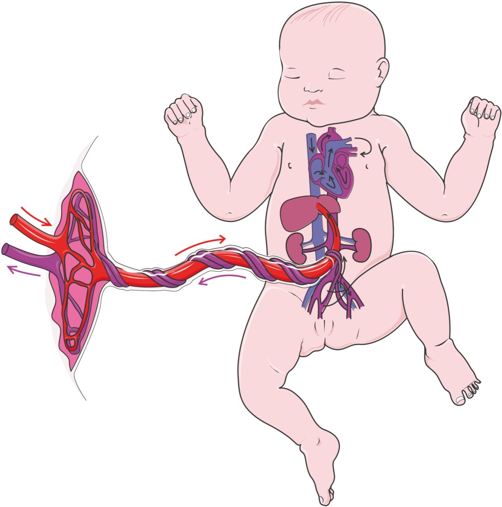
The umbilical cord connects the placenta to the fetus. The fetus develops directly from the placenta. The cord attaches to the fetus’s abdomen. This cord has three important blood vessels inside it :
- The umbilical vein is responsible for carrying oxygenated and nutrient-rich blood from the placenta to the fetus.
- the two umbilical arteries carry deoxygenated blood and waste products from the fetus to the placenta. This is for reoxygenation and elimination. The umbilical arteries come from the baby’s internal iliac arteries. The cord is filled with a soft, jelly-like material called Wharton’s jelly. This material cushions the blood vessels. It protects them from getting twisted or squeezed.
Fetal Shunts:
The fetal circulatory system utilizes three specialized shunts. These shunts ensure efficient oxygen delivery to the developing tissues and organs. They allow blood to bypass the non-functional lungs and the not fully mature liver.
The ductus venosus is a vital fetal blood vessel that connects the umbilical tone directly to the inferior vena cava. It primarily allows much of the largely oxygenated blood to bypass the liver. This blood is returning from the placenta via the umbilical tone. This allows it to enter the systemic rotation more directly. This shunting medium ensures that the oxygen-rich blood reaches the fetal heart quickly. It then reaches the brain with minimum detention. This supports their critical development. The inflow of blood through the ductus venosus is regulated by a sphincter. It allows some control over the quantum of blood that bypasses the liver.
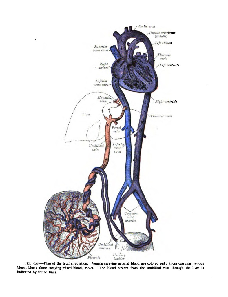
A small opening between the right and left atria called as foramen ovale is present in the fetal heart. It lets blood flow directly from the right atrium to the left, skipping the right ventricle and the unused lungs. This enables oxygenated blood from the placenta to reach the rest of the fetus’s body efficiently. The pressure in the right atrium is higher than in the left. This difference pushes most of the oxygen-rich blood from the inferior vena cava through the foramen ovale. It enters the left atrium after this. From there, the blood flows into the left ventricle. It is then pumped into the aorta. This process supplies oxygen to the body. It includes important organs like the brain and heart. This bypass is essential because it ensures that oxygenated blood from the placenta is preferentially directed to the systemic circulation. It mainly supports the upper body and brain. This happens without the need to circulate through the unused lungs.
The ductus arteriosus is a short and wide blood vessel in the fetal heart. It connects the pulmonary artery to the descending aorta. Its main role is to allow most of the blood from the right ventricle to bypass the fetal lungs. These lungs are non-functioning, filled with fluid, and not yet used for breathing. Instead, the blood is directed straight into the systemic circulation, helping deliver oxygen and nutrients to the growing body. The fetal lungs are filled with amniotic fluid. They have a very high vascular resistance. Roughly 90% of the blood flow in the pulmonary box is diverted through the ductus arteriosus into the descending aorta. This medium protects the immature lungs from circulatory load. It also allows the right ventricle to strengthen for its full function after birth.
The path of oxygenated and deoxygenated blood :
Oxygenated blood, rich in essential nutrients, flows from the placenta to the fetus through the umbilical vein. This blood provides the necessary oxygen and nourishment for the fetus’s growth and development. It also supports the fetus’s overall health while inside the womb. After entering the fetus at the umbilicus (belly button), the umbilical vein moves towards the liver.
There it divides into branches. The ductus venosus lets the oxygenated blood bypass the liver. It flows directly toward the fetal heart. This allows the blood to quickly join the inferior vena cava (IVC). The IVC is a major vein that carries blood toward the heart. Meanwhile, a smaller portion of the umbilical blood flows through the liver tissue. This nourishes it with oxygen and nutrients. After being nourished, it also joins the circulation.
Inside the IVC, the oxygen-rich blood from the placenta mixes with blood from the lower parts of the fetus’s body. This blood is deoxygenated. These include the legs and abdominal organs. This mixed blood flows into the right atrium of the fetal heart. Because of pressure differences, most of it passes through a special opening called the foramen ovale into the left atrium.
This shortcut helps more oxygen-rich blood reach the left side of the heart quickly. From there, the blood moves into the left ventricle. The left ventricle then pumps this blood into the ascending aorta. This large artery carries blood upward toward the brain, heart muscle (via coronary arteries), head, neck, and upper limbs. These organs, especially the brain and heart, require a rich supply of oxygen for their rapid growth and development.
Some blood continues down the descending aorta. It supplies the rest of the fetus’s body, including the abdomen and lower limbs. This unique circulation pattern ensures the most oxygenated blood is delivered first. This priority delivery goes to the most critical organs—the brain and heart. This delivery supports their high metabolic needs during fetal development.
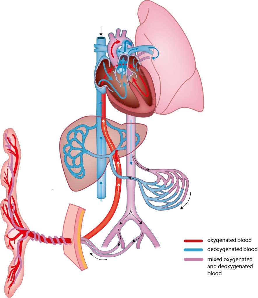
Fetal Circulation of Deoxygenated Blood :
1. Fetal Tissues – Oxygen used for metabolism – Blood becomes deoxygenated (high in CO2 and waste)
↓
2. Upper Body (brain, head, neck, arms) – Deoxygenated blood returns via Superior Vena Cava (SVC)
↓
3. Lower Body (abdomen, legs, liver) – Deoxygenated blood returns via the Inferior Vena Cava (IVC) – Mixed with oxygenated blood from the placenta via Ductus Venosus (implied)
↓
4. Right Atrium (RA) – Receives blood from both SVC and IVC
↓
5. Right Ventricle (RV) – Pumps blood into the Pulmonary Trunk
↓
6. Pulmonary Artery Divides – Small Portion: Blood flows to developing lungs (non-functional for gas exchange) – Majority: Bypasses lungs Via Ductus Arteriosus
↓
7. Ductus Arteriosus – Connects the pulmonary artery to the descending Aorta
↓
8. Descending Aorta – Distributes deoxygenated blood to Lower Body and internal iliac arteries
↓
9. Internal Iliac Arteries – Give rise to umbilical Arteries (2)
↓
10. Umbilical Arteries (2) – Carry deoxygenated, waste-laden blood back to the placenta
↓
11. Placenta – Fetal blood releases CO2 and waste to Maternal blood -Fetal blood absorbs oxygen and nutrients – Supports fetal growth and development
↓
12. Umbilical Vein – Begins the oxygenated blood pathway – Carries oxygenated blood from placenta towards fetal liver/IVC – Mixes with deoxygenated blood in IVC – Oxygenated blood goes to fetal liver and then IVC, or directly to IVC via Ductus Venosus (implied)
Additional Pathway (Implied): – Foramen Ovale: RA to LA (Left Atrium) – LA to LV (Left Ventricle) – LV to Ascending Aorta – Ascending Aorta supplies Upper Body (brain, head, neck, arms)
Circulatory Changes at Birth: Transition to Neonatal Circulation:
The moment of birth triggers a series of dramatic changes in the infant’s circulatory system. These changes happen rapidly. The system transitions from the fetal pattern to the independent, adult-like configuration.
- Clamping the umbilical cord removes the low-resistance placenta, making the baby’s circulation more dependent on its own heart.
- Increases pressure in body circulation
- First Breath:○ Lungs expand.
- Resistance in the lung vessels drops.
- More Oxygen:○ Causes the ductus arteriosus to close.
- Pressure Shift in the Heart:○ Left atrium pressure rises, right atrium pressure drops.
- Foramen ovale closes.
- Ductus Venosus Closes:○ Blood now flows through the liver
- Final Changes:○ All shunts become ligaments
- Lungs and liver now handle oxygen exchange and metabolism.
Potential Complications and Abnormalities :
Persistent Fetal Circulation (PFC):
○ This condition is also referred to as persistent pulmonary hypertension of newborn.
○ Blood keeps skipping the lungs, causing low oxygen levels.
○ Can be due to underdeveloped lungs, certain illnesses, or stress.
Congenital Heart Defects:
○ Structural issues in the heart that interfere with normal circulation.
○ Examples: ASD, VSD, PDA, Tetralogy of Fallot.
Patent Ductus Arteriosus (PDA):
○ Ductus arteriosus stays open after birth.
○ It causes more blood to flow into the lungs.
Placental Problems:
○ When the placenta doesn’t work well, it can reduce the oxygen and nutrients the baby gets.
○ May lead to growth problems or abnormal blood flow.
Medications and Other Conditions:
○ Some drugs taken during pregnancy (like NSAIDs) can affect fetal circulation.
○ Abnormal readings in prenatal ultrasounds can also show risks.
Conclusion :
Fetal circulation is a unique system that helps the baby grow and develop in the womb. The lungs and liver aren’t fully active before birth. Therefore, the placenta takes over their roles by delivering oxygen and nutrients. This system uses three unique shunts—the ductus venosus, foramen ovale, and ductus arteriosus. These shunts direct blood efficiently. This ensures the brain and heart receive the most oxygen-rich blood. Oxygen-rich blood from the placenta travels through the umbilical vein. It skips most of the liver via the ductus venosus. Then, it enters the heart. It then flows through the foramen ovale into the left side of the heart, which pumps it to vital organs. Blood from the right ventricle bypasses the non-working lungs through the ductus arteriosus and flows into the aorta. After birth, this system quickly changes. The lungs begin working. The placenta is no longer needed. The shunts close, allowing normal blood flow through the lungs and liver. Understanding this process is key. Recognizing and treating heart or circulation problems early are critical. Simple explanations make this complex system easier to appreciate.


Leave a comment